INTERCENTIONAL CARDIOLOGY


As with all other diseases, the early diagnosis of cardiac diseases is vitally important for successful treatment. Regular check-ups and close monitoring of the health of your heart is the first condition of early diagnosis. Acıbadem Heart Care Centers provide diagnostics and treatment using state-of-the-art equipment.
ECG: Electrocardiography (ECG) is a device that records the electrical activity of the heart to examine the cardiac muscle and its functioning. It is used for fast assessments, especially in emergencies.
ECG is an important tool in the diagnosis of cardiovascular diseases, structural anomalies, and arrhythmias. ECG monitoring and interpretation can be performed quickly.
The Effort ECG Test:This is an exercise test performed on a treadmill following a systematic, specific protocol. It is based on the interpretation of ECG recordings received via electrodes placed on the chest while exercising. The test usually lasts 5 to 10 minutes, but varies based on the patient’s age and condition.
It is a test that monitors the functioning pattern of the heart under effort and is used to identify embolisms that normally do not display symptoms in daily life.
Interpretation of the Effort EKG test for diagnosis should be done by experienced doctors to avoid misinterpretations about some other diseases with similar findings.
Echocardiography:Echocardiography is a diagnostic and investigative tool that allows examining the structure, pathology and functions of the heart by using ultrasonic sound waves.
It is possible to examine the movements and cavity of the ventricle wall, the growth of the cardiac muscle, and cardiac valves using echocardiography. It also makes it possible to observe the structure and functionality of implanted artificial valves. Virtually all congenital cardiac diseases are diagnosed using this method.
It has no harmful side effects on the patients and can be used easily. The patient feels no pain during the procedure.
Holter Monitorization: A Holter monitor is used to monitor the cardiac rhythm or blood pressure of the patient. Separate devices the size of cell phones are used for EKG records and blood pressure measurements. The devices are affixed to the body of the patient for usually 24 hours or more. The devices measure cardiac rhythm and blood pressure continuously.
It is usually used to monitor the cardiac rhythm and blood pressure of the patient in daily life. Physicians use this test upon suspicion of an abnormal cardiac rhythm or an imbalance in blood pressure.
Trans-Telephonic Monitor: A recording device, similar to the Holter device, is attached to the patient to monitor cardiac functions.
Under normal circumstances, Holter devices can remain on the patient for two to three days. However, in patients who only rarely feel any discomfort, symptoms may not occur during the time the Holter device is carried. In such cases, a telemedicine device that operates trans-telephonically can be used.
Stress Echocardiography Echocardiography taken during periods of rest determines the width of the cardiac cavity, malfunctions in the movement of the wall, and contraction functions of the heart. It can help to indirectly diagnose coronary artery disease. It also helps to identify other conditions such as cardiomyopathy that compliment other valve diseases, cardiac membrane infections, a tear in the aorta, and excessive thickening of the heart that could cause chest pains and difficulty in breathing.
Stress echocardiography can be used in conjunction with the effort ECG to identify the location of vascular disease.
Myocardium Perfusion Scintigraphy: Myocardium perfusion scintigraphy is used mainly to identify any problems in blood accumulation in the cardiac muscle. It provides information about the blood build-up of the heart under two different conditions, one under stress (for instance, during exercise) and the other, in rest.
Myocardium scintigraphy can be used to identify serious coronary artery diseases. Its diagnostic sensitivity and precision in the diagnosis of severe vascular diseases is around 90 percent. Findings obtained during the test also provide information on the mortality risk, cardiac functions, and advanced cardiac failure of the patient, and data vital in making a decision about the correct course of treatment.
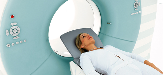
Flash CT: Flash CT is a radiological method of diagnosis that creates a cross-section image of the examined area using X-rays.
As a radiological method of diagnosis, the Flash CT is able to provide images of all the parts of the body, particularly cardiac and pulmonary scans.
The heart can be scanned in 250 milliseconds. When compared with single tube and single detector systems, it provides images in half the time. It allows the heart to be scanned in 250 milliseconds, with a 99% accuracy rate. (a quarter of the time of a heartbeat). Thus, although even in cases in which the heart rate of the patient is over 100 beats per minute, there arises no need to slow the heart down with medication. . Flash CT is a scanning tool that emits the lowest level of radiation in the market. Cardiac scanning can be completed with 80 percent less radiation. It can be used in routine procedures as a non-invasive cardiologic diagnostic method.
Cardiac MRI Test: The MRI Test provides valuable information on congenital cardiac diseases and cardiac cavities and enables detailed assessments of structures of the main arteries entering and exiting the heart. It supplements echocardiography findings without adversely affecting the patient.It provides detailed information in the assessment of cardiovascular constrictions, the extent of the effect of a heart attack on cardiac muscles, and the condition of the heart in preserving its vitality and functionality. The MRI test has the highest level of diagnostic sensitivity in the assessment of cardiac muscle diseases and masses in the heart.
Contrary to traditional imaging methods, the MRI does not contain radiation and ultrasound waves. The accurate images of the organs are displayed using physiological parameters.
PET CT: The PET CT is a cardiac examination based on scintigraphy. This method is utilized to observe vitality retention of the heart. It is used mainly to obtain detailed information on the function and vitality of cardiac cells, giving accurate results on the vitality of cardiac tissues. It provides guidance in determining whether bypass surgery would be beneficial for a high risk patient.

